
Beta · design partners
Annotate together.
Annotate together.
Find anything you've marked.
Your team marks cells, structures and regions on one shared layer, and labels them with cell types and other terms from your own lists or published ontologies. Search any value and the image shows exactly what you found. No shader code, no files passed around.
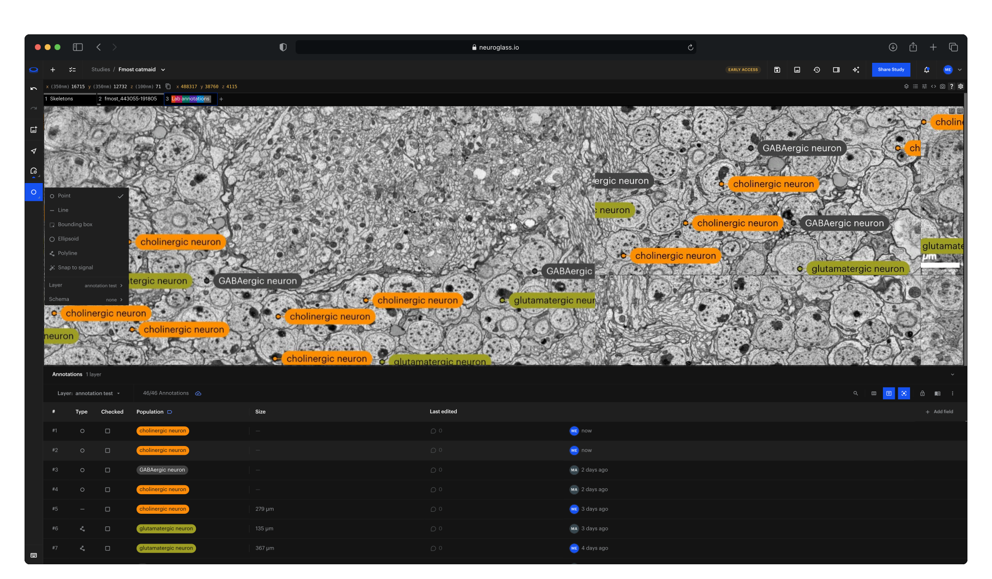
One shared layer
The whole team, and the specialists you invite from other labs, annotate the same copy in the browser. Changes show up for everyone as they are saved.
Values everyone agrees on
Fields are defined once, as lists, numbers or yes/no, or bound to published ontologies, so a label means the same thing to every reader, every lab and every script.
Found without code
Search by any value and the image shows exactly those annotations. Nobody writes shader code to answer a question.
Why it matters
No more links, files and spreadsheets
Most teams keep annotations wherever they were made: inside a Neuroglancer link that grows too long to share, in precomputed files that need their own tooling, in a spreadsheet next to the image, or in a desktop tool only one person can open. The work is there, but the team can't search it, and colleagues in other labs can't add to it.
No more shader code
Search any value and the image shows exactly the annotations that match, ready to review.
No more overwritten work
Everyone edits one shared layer: a clashing save is refused, and each annotation shows who changed it last, and when.
No more free-text labels
Cell types and other values come from your own lists or published ontologies, so they mean the same thing to every reader, every lab and every script.
What you can do
From the first mark to a layer the team trusts
Set up · Fields
Agree on what to record
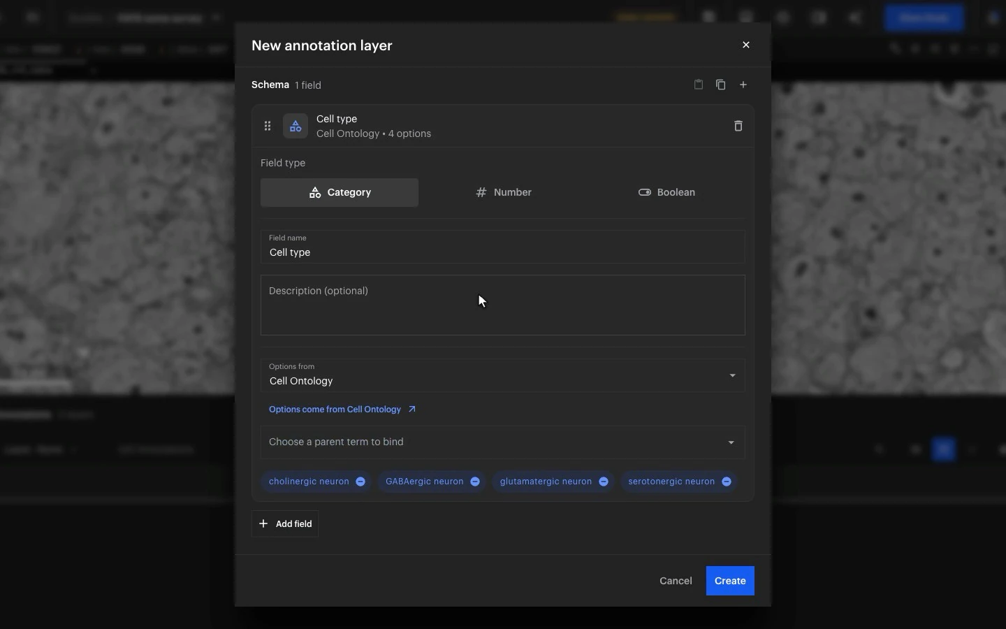
Before anyone marks a cell, decide what every annotation records: a category, a number or a yes/no. A category's options can be a list you type, or the terms under a parent term in the Cell Ontology, Uberon or an Allen atlas, like the neuron types here, so everyone picks from the same values.
Annotate · Mark
Mark cells and structures on the image
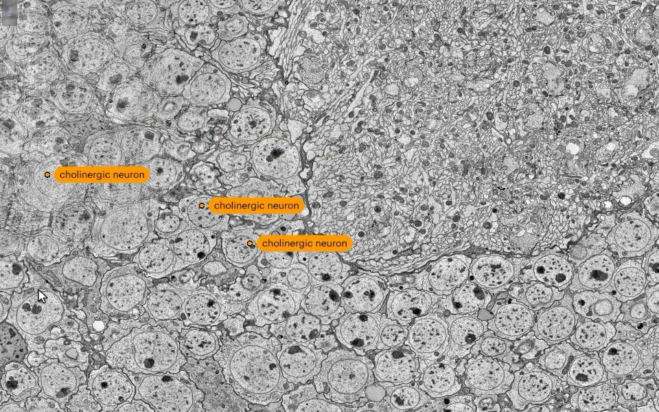
Place points, lines, boxes and ellipsoids on volumes of any size, streamed from your own storage, or trace a structure with a line that snaps to the signal. Labels sit on the image, and a label's info button shows what its term means, straight from the ontology.
Annotate · Share
Annotate alongside your colleagues
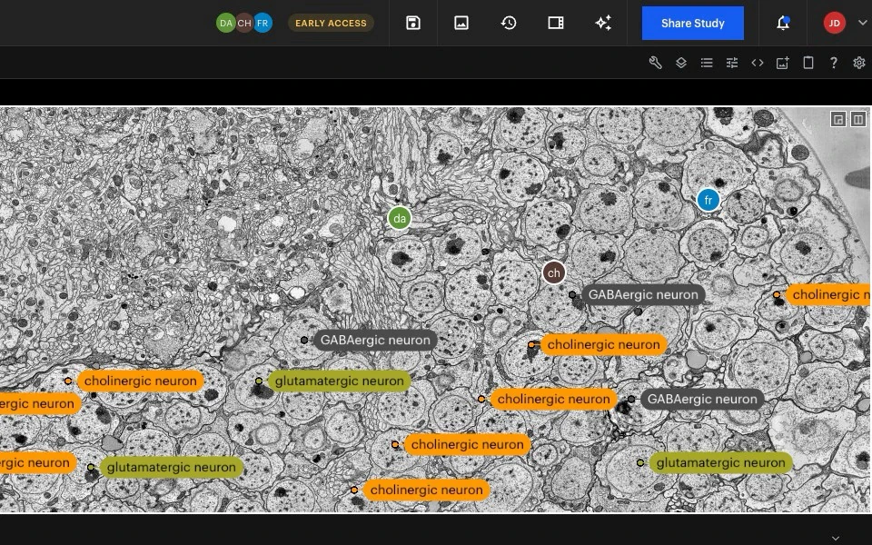
Share the study, and colleagues or specialists from other labs work on the same layer with you. You see who is in the study and where each of them is looking, and their changes appear as they're saved. If two people save the same annotation at once, the later save is refused and shows the newer values, so nobody overwrites a colleague.
Review · Filter
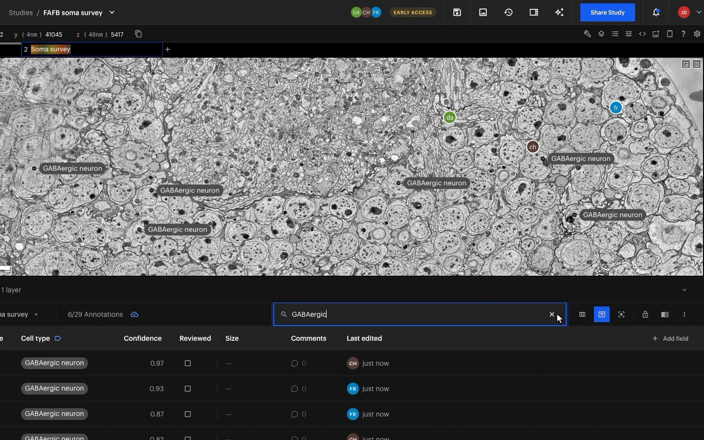
See only the cells that match
Filter the annotation table by any value and the image shows only the annotations that match. Type GABAergic and the other 23 cells drop out of view, leaving the six to look at, with no shader code to write. Clear the filter and they all come back.
Review · Check
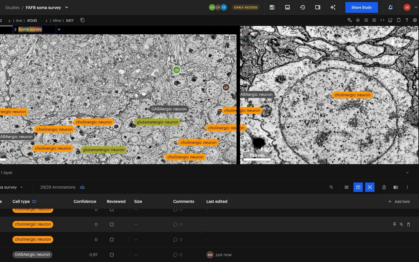
Check them one by one, up close
Click a row to go to its annotation. Focus View adds a close-up beside the overview that follows the row under your pointer, so you can check cell after cell without losing your place.
Review · Discuss
Ask a colleague, right on the cell
Start a thread on any annotation and @mention a colleague, who gets a notification. The comment stays pinned to the cell, so whoever opens it sees the question and the cell together.
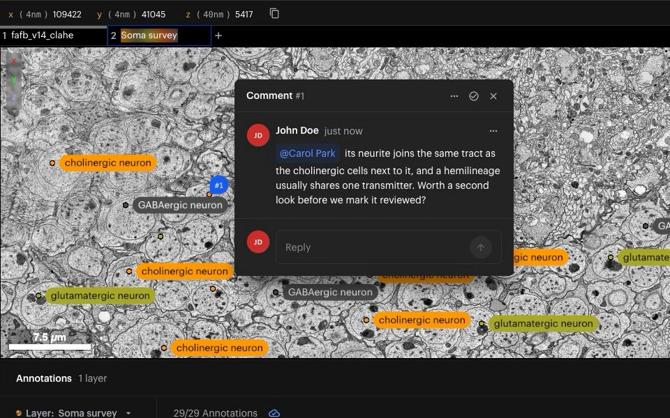
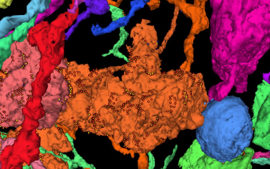
Images: FAFB, the full adult fly brain (Zheng et al., Cell 2018), with example annotations.
How teams use it
From screening plates to brain volumes

Screening plates
Record the compound, concentration and cell line of every field of view on the plate itself, then find the wells by their values and look straight at their cells.

Brain slices
Mark cortical layers, landmarks and recorded cells on slice overviews with terms from the Allen atlases, alongside colleagues, and let the lab's software read the result.

Volume surveys
Label cell bodies by cell type, synapses or regions across electron-microscopy and light-sheet volumes, share the work with specialists from other labs, and see what has been checked.
Built on Neuroglancer
Bring the annotations you already have
Collaborative Annotation works with the formats your team already uses, and your images stay where they are.
Copy precomputed annotation layers, and layers saved inside a Neuroglancer view, into a shared layer the team can edit
Read and write annotations from your own scripts and pipelines through the API
Keep streaming images from your own buckets: nothing is copied
Run it on our cloud or inside your own deployment, like the rest of NeuroGlass
Design Partner Program
Built hand-in-hand with our design partners
Collaborative Annotation is in production with a small group of labs and companies. Partners get early access, shape what we build next, and a direct line to our team.
Frequently Asked Questions
Can't find an answer? Contact us
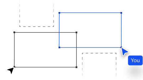
Which kinds of annotation can we make?
Can we use our own vocabulary, or a published ontology?
We already have annotations in Neuroglancer. Can we bring them?
Who can see and change our annotations?
What happens if two people edit the same annotation?
Does our image data leave our storage?
How is the module priced?
Modules
Explore the other modules
Beta · design partners
Skeleton Tracing
Light-sheet
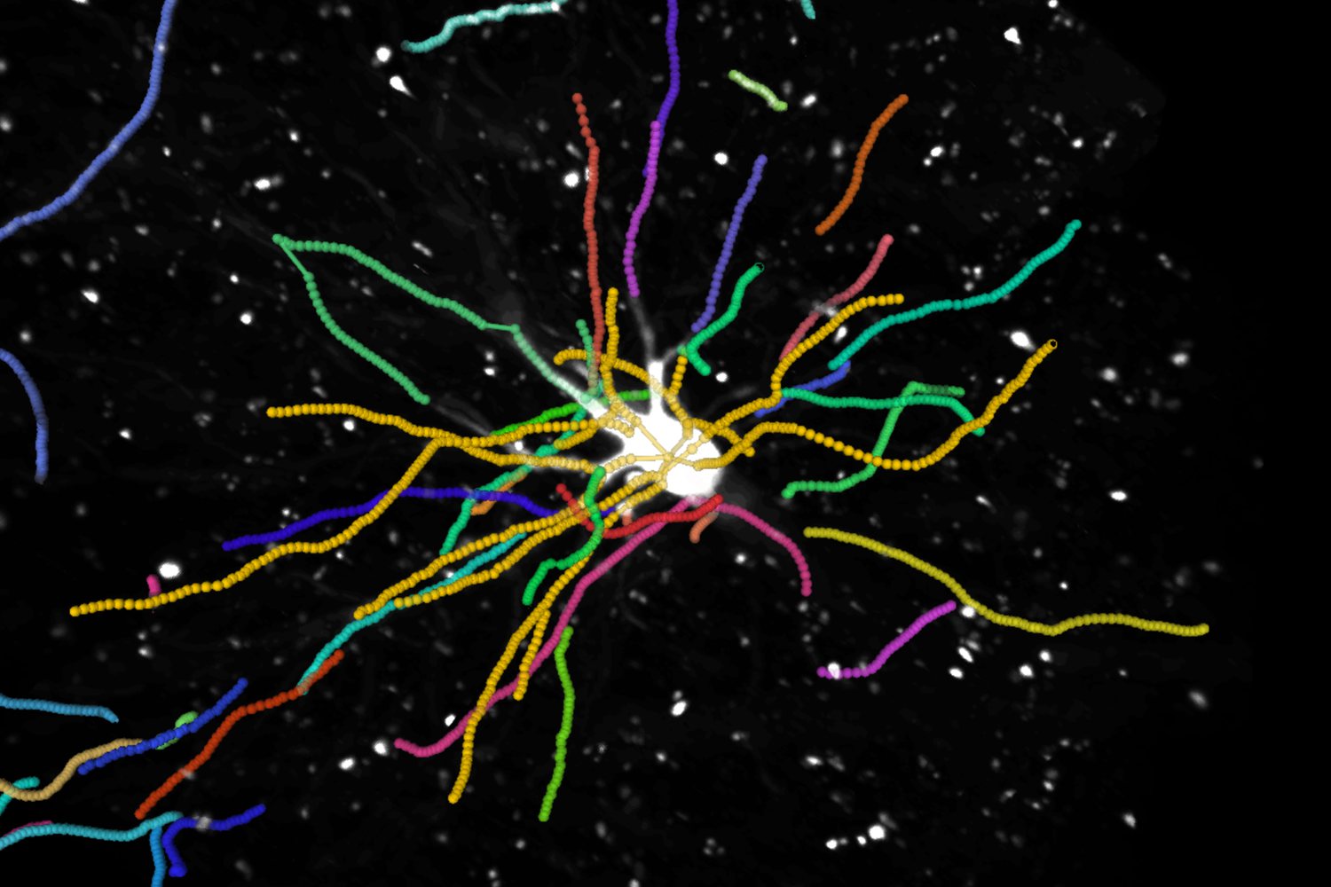
Trace and proofread neurons in the browser with your whole team, one task per neuron, and review each one in the same view.
Coming soon · design partners
AI Segmentation
Fluorescence · LSFM · EM
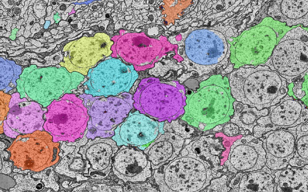
Kick off automated segmentation over a region of interest, then monitor and approve the results.
Coming soon · design partners
High-Content Screening
2D assays · organoids
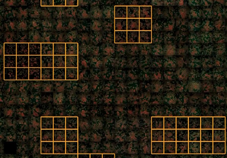
Review, QC and confirm hits at plate scale, with your feature tables and masks overlaid on the wells.
Coming soon · design partners
EM Reconstruction
EM · Tomography
Build and proofread ground-truth reconstructions, with a dedicated proofreading task type and review UX.


