The collaborative platform for
large-scale volumetric imaging.

Explore, annotate and collaborate on terabyte-scale imaging volumes

Trusted by teams at
Allen Institute logo Calico logo BossDB logo Translucence Biosystems logo MIT logo Massachusetts General Hospital logo Harvard Medical School logo Harvard University logo University of Edinburgh logo E11 Bio logo Brigham & Women's Hospital logo University College London logo Columbia University logo Duke University logo Weill Cornell Medicine logo Université Claude Bernard Lyon logo Universidad San Sebastián logo
Work together. Discover more.

Collaboration should not be hard

Discuss, share and work together on the same Study — comments, permissions and live sessions, built in.

Built to scale

Shareable Studies created

19,000+

reproducible Glances captured

21,000+

Figures created

26,000+

Modules

Additional Modules for your most challenging workflows

Turn any study into a reconstruction and analysis workspace. Modules run on dedicated cluster backend services, on top of a Team or Enterprise plan. Your data never leaves your own storage.
01 / 05
Beta · design partners

Skeleton Tracing

Light-sheet
Trace and proofread neurons in the browser with your whole team, one task per neuron, and review each one in the same view. In production with design partners today.
02 / 05
Beta · design partners

Collaborative Annotation

All modalities
Annotate together on one shared layer, with values from your own lists or published ontologies, and find any annotation by its values, without code. In production with design partners.
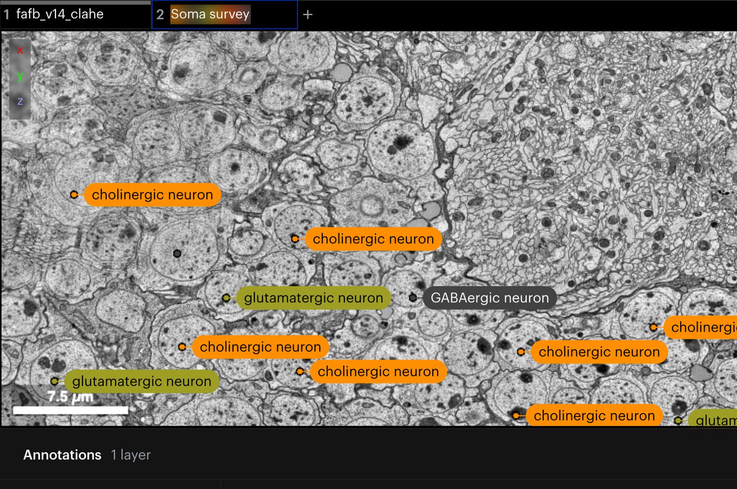
Cell bodies in a fly brain electron-microscopy volume, each labelled with its cell type from the Cell Ontology, above the annotation table.
03 / 05
Coming soon · design partners

AI Segmentation

Fluorescence · Light-sheet (LSFM) · Electron microscopy
Kick off automated, multi-channel segmentation over a region of interest, then review, correct and approve the results in-platform. A real pilot in progress with our design partners — not yet generally available.
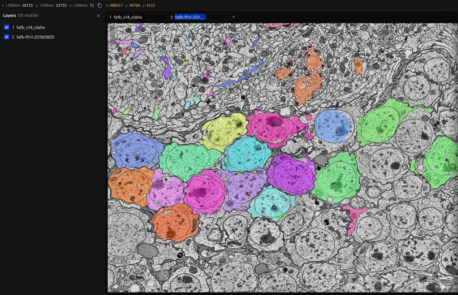
NeuroGlass with a full-screen 2D electron-microscopy view and a cell segmentation layered on top.
04 / 05
Coming soon · design partners

High-Content Screening

High-content screening · 2D assays · organoids
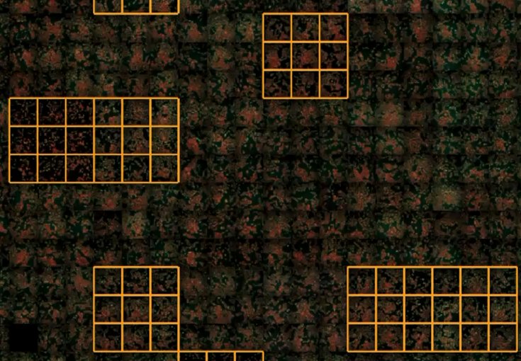
Review hits at plate scale. Overlay tables and masks on wells, standardize views, flag artifacts, and capture decisions as reproducible Glances. We provide the review layer on your screen, developed with design partners.
A high-content plate in NeuroGlass, with groups of annotated wells outlined.
05 / 05
Coming soon · design partners

EM Reconstruction

Electron microscopy · Tomography
Build and proofread ground-truth reconstructions of electron-microscopy and tomogram volumes, with task assignment and review. In active development with our design partners.
A 2D electron-microscopy section beside the 3D reconstruction of its segmented neurons.

From cells to whole brains

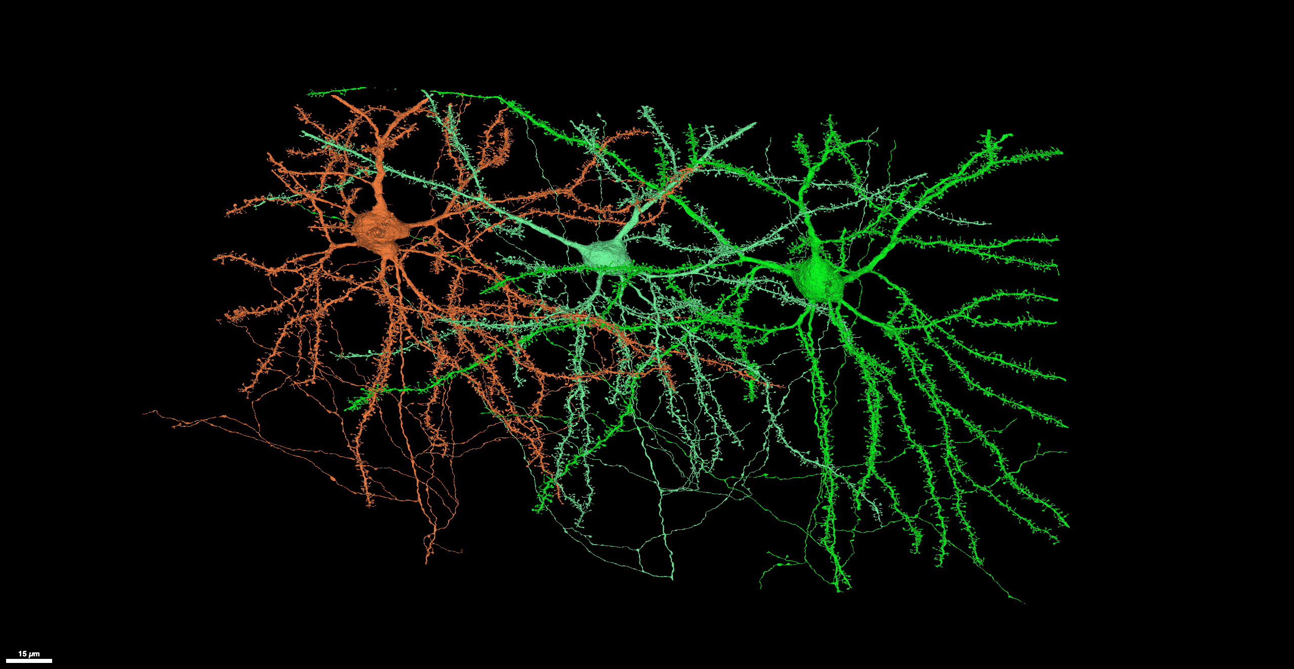
Three colored neurons with branching dendrites visualized against a black background.

Connectomics

Explore neurons across EM volumes
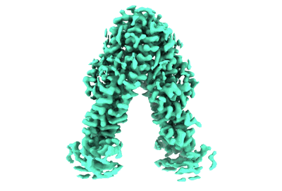
3D cryo-electron microscopy density map of a molecular structure with a triangular arch shape in teal color on a white background.

Cryo-EM/ET

Overlay particle picks on tomograms
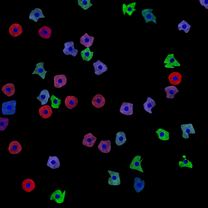
Fluorescently stained cells in red, green, and blue scattered on a black background, with nuclei distinctly highlighted in blue.

Expansion Microscopy

Review expanded tissue across channels
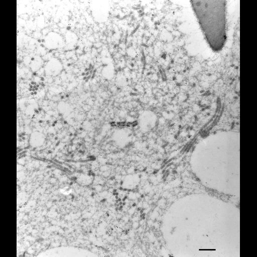
High-resolution grayscale electron microscope image showing a dense network of cellular structures including vesicles, microtubules, and other organelles within a cell.

High-Res Cellular Imaging

Navigate organelles in 3D
Close-up of a textured surface featuring a dense pattern of multicolored speckles primarily in blue, orange, and purple hues.

Spatial Transcriptomics

Map gene expression onto tissue
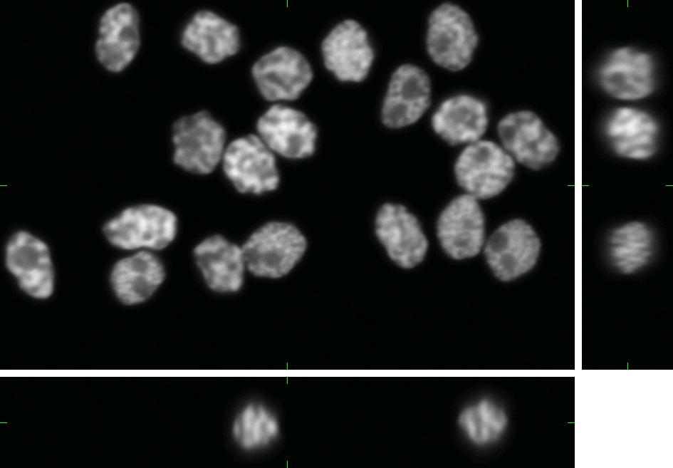
Microscopic grayscale image showing multiple cell nuclei with varied shapes and detailed internal structures on a black background.

Developmental Imaging

Compare samples and timepoints
Microscopic image showing green and purple molecular structures on a dark background.

Molecular Imaging

Overlay molecular channels in context

Electron Microscopy

Fluorescence Microscopy

Attributions

What's new

Nov 23–24, 2026 · Symposium

Meet us at the Crick BioImage Analysis Symposium in London

Nov 14–18, 2026 · Conference

Meet us at Neuroscience 2026 (SfN) in Washington, DC

Nov 2–4, 2026 · Conference

Meet us at the SBI2 annual conference in Boston

Oct 26–29, 2026 · Conference

Meet us at the Global BioImaging Exchange of Experience at EMBL Heidelberg

Jun 10, 2026 · Press Release

Allen Institute using NeuroGlass by MetaCell to support large-scale brain imaging workflows

May 3, 2026 · Webinar

The Collaborative Platform for Volumetric Imaging Datasets

Your data, protected by design.

Research securely. Collaborate confidently.

We take your data security seriously — that’s why we ensure it never leaves its trusted home. SSO, role-based access, EU data residency, and on-prem deployment with no external cloud dependencies.
See security & deployment

Secure hosting

Whether deployed in your institution or on MetaCell Cloud, your research data never leaves your servers.

GDPR compliance

Your information is safe with us. Visit the Privacy Terms to know more about how we treat personal information.

Secure Single sign-on (SSO)

Integrate NeuroGlass with your authentication provider for easy access.