
Drug screening
Review imaging evidence together,
from screens to whole organs
Images, analysis outputs and expert decisions in one shared workspace, on your own cloud or on‑prem.
Trusted by leading institutions worldwide













why imaging discovery stalls
Image-based discovery is fragmented across tools, experiments and scales
A single result crosses analysis software, spreadsheets, screenshots and email before anyone can act on it — and the trail back to the source image is lost along the way.
No path back to the image
A statistical hit is hard to trace to the exact well, field or structure it came from.
AI outputs go unchecked
Model results are hard to review together and reproduce across a team.
Context evaporates
Decisions, comments and rationale are lost between stages, tools and collaborators.
one review, start to finish
From hit to decision, in one place
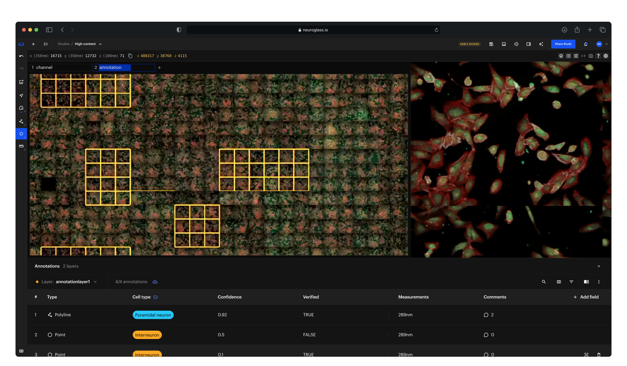
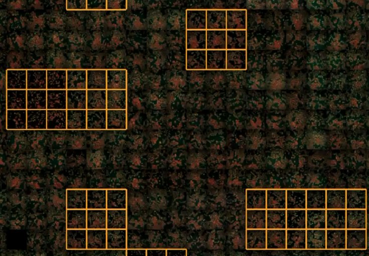
Spot the hit on the plate, open its wells, and save the call as a Glance anyone on the team can reopen.
1 · Spot the hit
Scan the whole plate in one view, with annotated wells outlined where they sit.
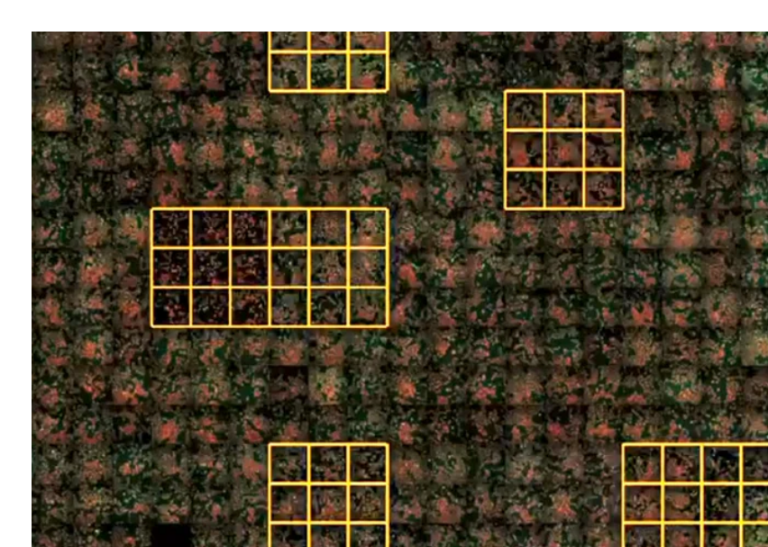
2 · Open the wells
Zoom from the plate into any well at full resolution, across every channel.
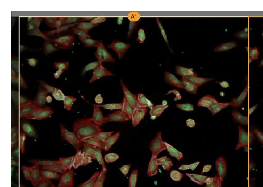
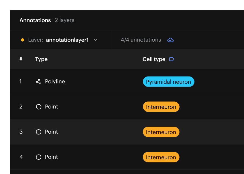
3 · Record the call
Annotate each call with its condition and well, then save the view as a Glance that reopens exactly as you left it.
one platform, every scale
From multi-well screening to whole-organ validation
The same collaboration, review and provenance layer spans every experimental scale — so image-based evidence stays connected as programmes move from cells to whole organs.
High-density screening
2D cellular assays
Many compounds across many wells, comparing treatment against control.
3D biology
Organoids & spheroids
Co-cultures, patient-derived models and other 3D phenotypes.
Whole-organ & neural
Cleared tissue to whole brain
Biodistribution and pathology mapping, with neuronal and axonal tracing across a whole brain.
Beneath every scale sits the same NeuroGlass layer — images, AI outputs and expert decisions together in one browser workspace, permissioned, versioned and reproducible.
Modules
Turn a screen into a reviewed, reproducible hit list
A module that layers onto your subscription and the analysis you already run. NeuroGlass is the review and QC layer on top of your screen — not another analysis engine.
Coming soon · design partners
Beta · design partners
High-Content Screening
High-content screening · 2D assays · organoids
Review, QC and confirm hits at plate scale — overlay your own analysis on the source wells and capture every advance-or-kill decision as a reproducible Glance. Runs on dedicated backend services, on your own storage.
Collaborative Annotation
All modalities
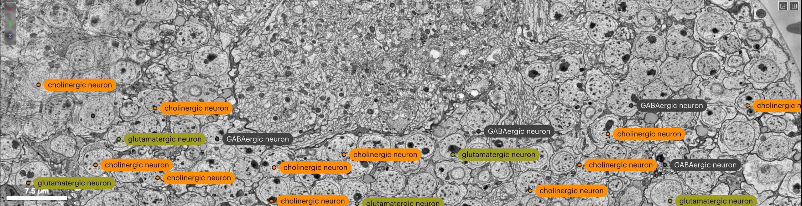
Annotate together on one shared layer, with values from your own lists or ontologies, and find any annotation by its values.
Bring your own analysis
Overlay existing feature tables and segmentation masks on the source wells at plate scale — CellProfiler, vendor software or your own models.
Review & QC hits
Standardize views with Lenses, flag edge effects and artifacts, and confirm or reject each hit with a spatial comment the whole team can see.
Every decision reproducible
Each advance-or-kill decision is tied to the exact well and view as a Glance — auditable and re-openable months later.
NeuroGlass for Pharma & Biotech
Enterprise-grade imaging for commercial R&D
Screening, spatial biology, translational and preclinical imaging — and longevity biotech — in one secure, compliant workspace. Your data stays in place; your teams stop wrestling with infrastructure and get back to the science.

SaaS or on-prem — your infrastructure
Deployment that fits your enterprise
Run it as managed SaaS, or deploy on-prem in Kubernetes on your own cloud — no external cloud dependencies.
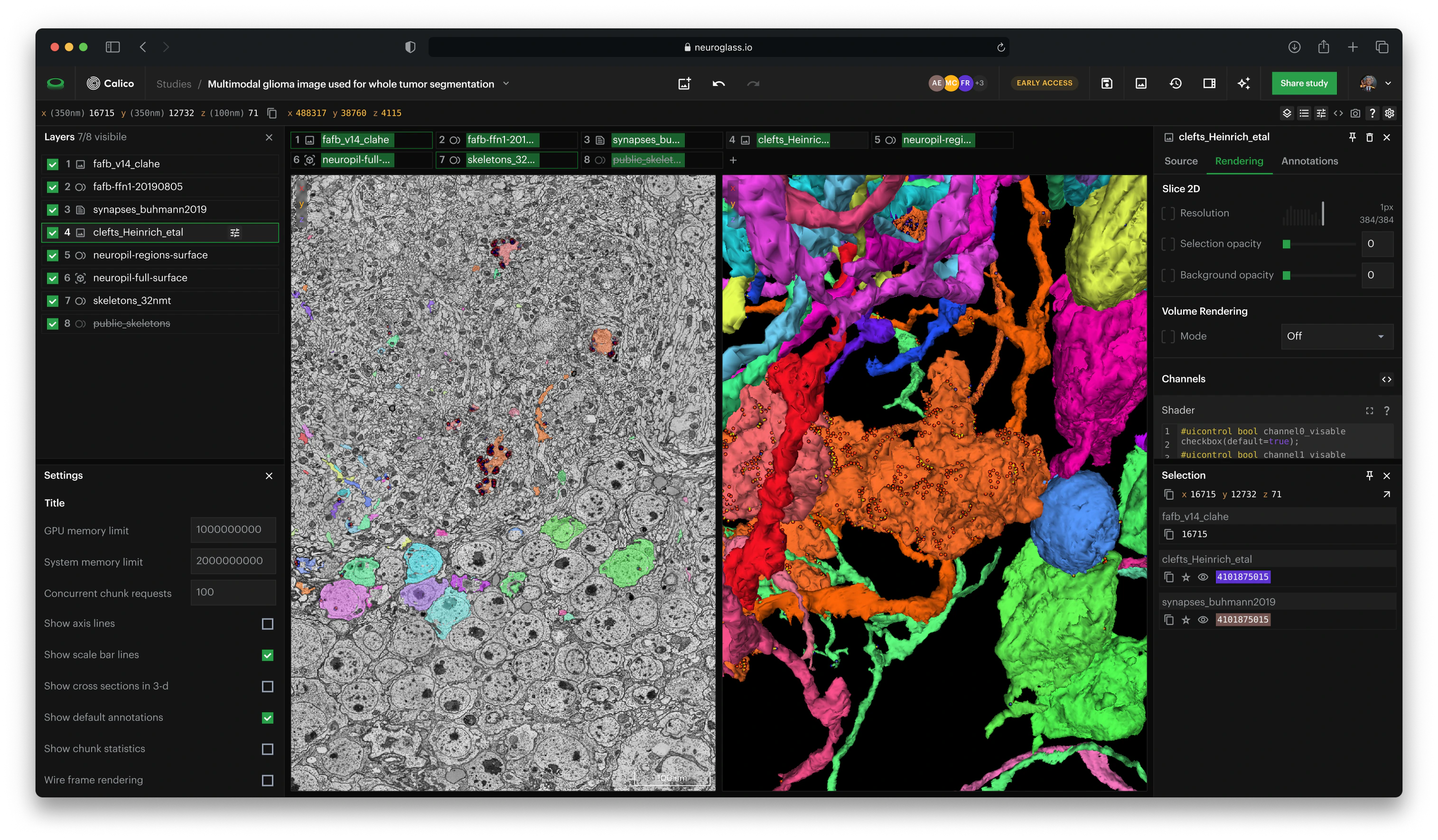
Attributions: Zheng et al., Cell 2018; Buhmann et al., Nature Methods 2021; Heinrich et al., arXiv 2018
bring your data, keep it in place
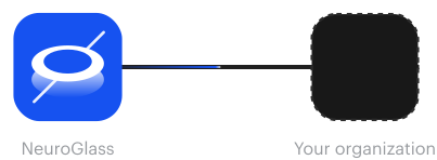
Your data never moves
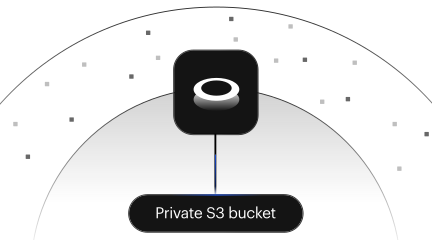
Point NeuroGlass at your existing private S3 buckets and stream directly into the browser — no copies, no migration, no data leaving your control.
Stream, don't copy
Terabyte-scale volumes render in the browser straight from your private buckets. No downloads, no duplicated datasets, no waiting on transfers.
Your storage, your rules
Keep data in your own account and region. NeuroGlass mediates access under your permissions — you retain full ownership and can revoke or delete at any time.
review where the data lives
Collaborate in the data, not around it
Discovery, translational and QC teams review the same images in one place, so every question, answer, and decision stays attached to the evidence — not buried in email, slides or PDFs.
Keep imaging data, annotations, quantitative outputs, and the exact Glance behind each finding in one place
Re-open any finding months later and see precisely what the scientist saw
Walk others through volumetric data in real time, or let them follow your demo remotely
Turn any question into a comment tied to a specific spatial point or ROI
Build the audit trail automatically as you work
screening & spatial at scale
Built for high-content and spatial pipelines
Bundle plates, cohorts and spatial panels into a Collection. Switch Lenses to change perspective.
Everything stays one click away in your dashboard.
Everything stays one click away in your dashboard.
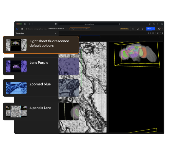
Standardize views
Lenses are parameterizable viewing templates that enforce consistent windowing, overlays, and rendering across every plate, cohort and scientist.
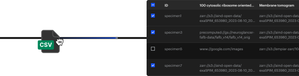
Bulk ingest
Onboard thousands of wells or samples in one pass with a CSV manifest, no bespoke scripts.
Compare anything
Swap datasets or swap settings. Dose-response, longitudinal and cross-cohort QC stop being engineering asks.
Secure. Compliant. Auditable.
Security and compliance built in
SSO and role-based access, a full audit trail, and deployment that keeps data in your own environment and region — designed to fit regulated pharma workflows.
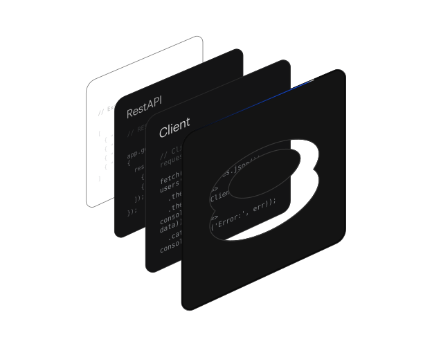
Visit API documentation

SSO & role-based access
Single sign-on and Google-Docs-style permissions via Keycloak, with a full activity audit trail across your organization
Built for regulated workflows
Deploy inside your own validated environment to support GLP and 21 CFR Part 11 workflows and GDPR, with EU data residency and data kept in your own region
On-prem, no external cloud
Run entirely in Kubernetes, on your own cloud or on-prem — no dependency on any external cloud service
Search across all your Data
Search Studies, Glances, and Collections and inside workspace state itself.
"The case where we saw that artifact" becomes findable.
"The case where we saw that artifact" becomes findable.
Example results from public datasets.
See it on your own data
The fastest way to evaluate NeuroGlass is to walk through your own imaging workflow end-to-end — ingest, explore, review, and reproduce — on your own data, in your own environment. Get the deployment checklist
Frequently Asked Questions
Can't find an answer? Contact us
What is NeuroGlass?
Which data formats does NeuroGlass support?
Do we have to upload or copy our data?
Where is our data, and who can see it?
Can NeuroGlass run inside our own environment?
Does NeuroGlass replace our image-analysis software?
Is the High-Content Screening module available today?
Allen Institute using NeuroGlass to support large-scale brain imaging workflows
Read the Press Release


